Early cornea development

As the optic cup (which will give rise to the retina) evaginates from the neural tube, it induces invagination of the lens vesicle from nearby ectoderm. The ectoderm remaining after lens invagination now overlays the optic cup and will eventually become the corneal epithelium. Meanwhile, NCCs migrate from the neural tube toward the developing eye. This migration is controlled by chemoattractant and repulsive factors in the surrounding microenvironment, some of which are produced by the lens. Once in the eye region, some NCCs invade the space between the lens and ectoderm to give rise to corneal stroma and endothelium, while other NCCs remain in the periocular region to give rise to the trabecular meshwork and other structures. Some molecules that control NCC invasion of the cornea have been identified, but mechanisms for NCC recruitment to the corneal fate are poorly understood. Learn more about our research on early corneal development.
Corneal Innervation

As the corneal layers are forming, other types of cells are also forming important structures in the developing eye. Nerves are recruited to the cornea, and they extend axons throughout the stroma and epithelium. The cornea is one of the most densely innervated tissues in the body, which makes it highly sensitive and helps to protect this vulnerable tissue from injury. Some molecules that control the innervation of the cornea have been identified, but the mechanisms for proper patterning of this innervation are still under investigation. Learn more about our research on corneal innervation.
Corneal Avascularity
Meanwhile, blood vessels are also recruited to the developing eye to supply this rapidly growing organ with oxygen and nutrients. However, blood vessels must be excluded from the adult cornea so that the cornea may be transparent. It is unclear at this juncture whether the cornea is avascular throughout development in all model organisms, or whether it is initially vascularized in the embryo and then later vessels are pruned. Understanding how the developing cornea initiates and maintains avascularity will provide important insights into inhibiting neovascularization of the adult cornea after infection or injury. Learn more about our research on corneal avascularity.
Corneal Wound Healing
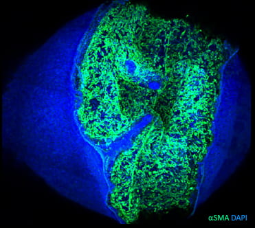 Finally, developmental programs that initially shape the cornea are also important in re-establishing corneal function after infection or injury. In the adult, the cornea has a limited capacity for wound healing, often forming a scar that obscures vision. Little is currently known about wound healing in the embryo, though there is hope that the embryonic cornea may have the capacity to heal scar-free. Co-opting the mechanisms by which scar-free wounds heal in the embryo has direct implications for fostering scar-free healing in the adult cornea. Learn more about our research on corneal wound healing.